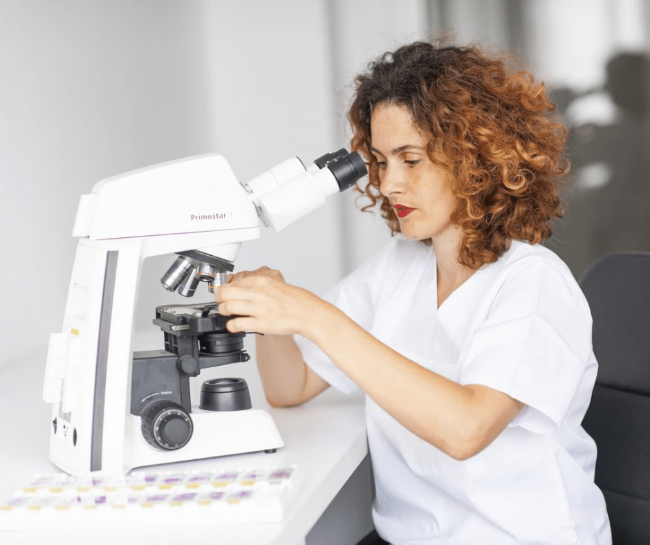
Histopathological speaking, endometriosis diagnosis is made when a lesion has endometrial type glands arranged in a fusiform, endometrial stroma. The histopathological diagnosis of endometriosis is made by a pathologist and consists of microscopic and macroscopic examination of tissues taken during surgery.
At the end of the surgery, certain endometriosis tissues from different locations such as ovaries, fallopian tubes, peritoneum, intestine are sent to a laboratory in special containers. Once they arrive in the laboratory, staff starts preparing, a process consisting of several stages, such as:
- Macroscopic examination and sampling of the necessary tissue fragments;
- Embedding in paraffin (wax);
- Microtomy and slide mounting;
- Staining and mounting on slides;
- Microscopic examination of slides;
- Drafting of the histopathological report by the medical anatomopathologist;
- Archiving.
All these stages are done with great care, by qualified personnel, following current protocols. At the end of this process, the pathologist’s findings will be included in a histopathology report.
What is the histopathology report?
A histopathological report is a medical document, written by a pathologist following the processing and interpretation of samples taken during surgery. The report contains the final diagnosis, which, when indicated, can be used to determine the treatment and prognosis of the condition.
The data that are included in the histopathological report are:
- Patient data
- Referral diagnosis
- Parts sent: ovary, fallopian tube, nodule, uterus, etc
- The referring physician
Apart from these data, the report must also contain:
- macroscopic description – what the specialist sees with the naked eye;
- microscopic description – what the doctor sees under the microscope. This stage is very important, and it’s dependent on the experience and knowledge of the anatomopathologist, as this one will confirm the diagnosis.
- diagnostic conclusion – to diagnose endometriosis, the examined tissue must contain both glands and stroma.
- the signature and initials of the pathologist.
Is diagnosis easy to make?
In the microscopic examination, the specialist identifies at least 2 features:
Endometrial glands formed from Müllerian epithelium. The specialist will also analyse whether the glands show degenerative atypia or metaplasia.
The endometrial stroma, usually containing a fine capillary network, may in turn undergo changes such as fibrosis or decidual changes.
There are also cases when the only component identifiable under the microscope is the stroma, and in this case we are dealing with a type of stromal endometriosis.
Although the histopathological diagnosis of endometriosis is easy to make; there may be difficulties resulting from changes in the endometriotic tissue over time. These changes may consist of alteration or absence of glands or stroma.
The stromal component can be altered by fibrosis, but also by myxoid changes and decidual changes. Also, the inflammatory and reactive changes that occur in endometriosis can complicate the histological results.
Another difficulty that can arise in histopathological diagnosis is when endometriosis tissues are associated with another condition, such as peritoneal leiomyomatosis or gliomatosis, resulting in a potentially confusing histological appearance.
The authors of a study carried out in Norway to find out the histological confirmation of endometriosis in various peritoneal lesions, show that of the 152 patients, endometriosis consisting of glands and stroma was diagnosed in only 78 patients. If the diagnostic criteria would have been extended to include stromal endometriosis, then 82 patients would have been diagnosed with endometriosis.
Immunohistochemistry, a plus in the diagnosis of endometriosis
In the case of specimens difficult to evaluate with classical histology, immunohistochemistry can be used to obtain a definite diagnosis. In such cases, the histopathological examination will be completed by the immunohistochemical examination of the paraffin-embedded tissue. The pathologist can use different immunohistochemical markers to confirm the diagnosis, such as CD10 – which will be positive in the endometrial stroma and ER, PR and PAX2 – which will be positive in the endometrial glands and endometrial stroma.

